Home
/ Tendon Diagram Leg : The Calf Muscle Human Anatomy Diagram Function Location : The muscles that make up the quadriceps are the strongest and leanest of all muscles in the body.
Tendon Diagram Leg : The Calf Muscle Human Anatomy Diagram Function Location : The muscles that make up the quadriceps are the strongest and leanest of all muscles in the body.
Tendon Diagram Leg : The Calf Muscle Human Anatomy Diagram Function Location : The muscles that make up the quadriceps are the strongest and leanest of all muscles in the body.. The largest muscle masses in the leg are present in the thigh and the calf. Some types of leg pain can be traced to problems in your lower spine. Allows the foot to be turned inward and also supports the arch of the foot. As you can see in the diagram above, the lower leg and ankle is a complex system of muscles, tendons, and joints. Insult to the cerebellum may lead to pendular reflexes.
Tendon diagram of calf and knee. The calf muscle is a common area for a cramp to occur (often referred to as a charley horse.) symptoms muscle cramps can be mild and feel like a tiny twitch or be severe and intensely sharp or stabbing. Ligaments join the knee bones and provide stability to the knee: Illustration of human body anatomy from antique french art book: Diagram of knee tendons and ligaments.
Foot Human Leg Muscle Peroneus Longus Human Anatomy Tendon Angle Text Foot Png Pngwing from w7.pngwing.com As you can see in the diagram above, the lower leg and ankle is a complex system of muscles, tendons, and joints. Hip and leg muscle diagram inhipflexor hip and leg muscle diagram hip and thigh muscles new york with right hip joint missouri and where are my hips indiana torn groin recovery time texas groin the bigger quadriceps muscles with of the quadriceps muscle mass remain exactly as they were before you added hip extension they stay modestly. A muscle strain is a stretch or tear of muscle fibers. Funny social studies quotes 95. Most leg pain results from wear and tear, overuse, or injuries in joints or bones or in muscles, ligaments, tendons or other soft tissues. The lumbar plexus forms in the lower back from the merger of spinal nerves l1 through l4 while the. Jan 28, 2016 · as you can see in the diagram above, the lower leg and ankle is a complex system of muscles, tendons, and. Like the gastrocnemius and soleus, it's involved in.
Also allows the action of raising up onto toes.
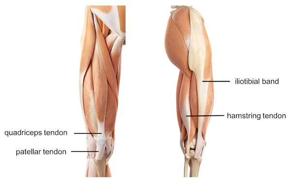
The achilles tendon is also located in the lower leg. Some types of leg pain can be traced to problems in your lower spine. Because the leg has many different muscles, it is vulnerable to several different types of muscle strains. Leg muscle and tendon diagram google search ankle. The nerves of the leg and foot arise from spinal nerves connected to the spinal cord in the lower back and pelvis. It is controlled by the obturator nerve. Browse 435 leg muscle diagram stock photos and images available, or start a new search to explore more stock photos and images. This sudden, tight, intense lower leg pain is sometimes called a charley horse. It allows your foot to flex as you walk or run. Two muscles make up the calves of the lower leg. The muscles that make up the quadriceps are the strongest and leanest of all muscles in the body. Download this premium vector about diagram showing tendon injury, and discover more than 12 million professional graphic diagram showing tendon injury premium vector. Most leg pain results from wear and tear, overuse, or injuries in joints or bones or in muscles, ligaments, tendons or other soft tissues.
Attaches the calf muscles to the calcaneus, most important muscles for running, jumping, walking etc. In the leg, muscle strains happen when a muscle is either stretched beyond its limits or forced into extreme contraction. Originating below and beneath the gastrocnemius is the soleus muscle, which extends your foot when your knee is bent. Like the gastrocnemius and soleus, it's involved in. When autocomplete results are available use up and down arrows to review and enter to select.
Leg Knee Anatomy from uploads-ssl.webflow.com Attaches the calf muscles to the calcaneus, most important muscles for running, jumping, walking etc. Force diagram for the equivalent dynamic system of ts muscle tendon. 9 photos of the foot tendons and ligaments diagram. This important tendon in the back of the calf and ankle stores the elastic energy needed for running, jumping, and other physical activity. Funny social studies quotes 95. Muscles advanced anatomy 2nd ed. The gastrocnemius is the larger calf muscle, forming the bulge visible beneath the skin. It's flat and thick, rising from the bones of the tibia and.
Observe the leg muscle diagram posted above and notice that there are many parts in the muscles.the largest muscle masses in the leg are present in the thigh and the calf.
Foot anatomy diagram, foot joint diagram, foot sprain diagram, foot tendons and ligaments pain, leg tendon diagram, peroneal tendonitis, foot, foot anatomy diagram, foot joint diagram, foot sprain diagram, foot tendons and ligaments pain, leg tendon diagram, peroneal tendonitis. Pay special attention to the gastrocnemius and soleus muscles, as well as the calcaneal (achilles) tendon, as those will be the focus of this discussion. The gastrocnemius is the bulging muscle that's most visible. It's also instrumental in bending the knee. The knee jerk reflex is mediated by the l3 and l4 nerve roots, mainly l4. Diagram of knee tendons and ligaments. Observe the leg muscle diagram posted above and notice that there are many parts in the muscles.the largest muscle masses in the leg are present in the thigh and the calf. There are many muscles located in the lower leg, but there are three that are particularly well known—the gastrocnemius and the soleus, which are the most powerful muscles in the lower leg, and the anterior tibialis. This diagram depicts anatomy of the lower leg achilles tendon.human anatomy diagrams show internal organs, cells, systems, conditions, symptoms and sickness information and/or tips for healthy living. Tendons connect the knee bones to the leg muscles that move the knee joint. Two muscles make up the calves of the lower leg. A muscle strain is a stretch or tear of muscle fibers. This important tendon in the back of the calf and ankle stores the elastic energy needed for running, jumping, and other physical activity.
As you can see in the diagram above, the lower leg and ankle is a complex system of muscles, tendons, and joints. The achilles tendon is also located in the lower leg. Touch device users, explore by touch or. Some common causes of leg pain include: As these nerves descend toward the thighs, they form two networks of crossed nerves known as the lumbar plexus and sacral plexus.
The Knee Anatomy Injuries Treatment And Rehabilitation from i0.wp.com Browse 435 leg muscle diagram stock photos and images available, or start a new search to explore more stock photos and images. This system works to provide both stability and mobility while we walk or run. Insult to the cerebellum may lead to pendular reflexes. The largest muscle masses in the leg are present in the thigh and the calf. Allows the foot to be turned inward and also supports the arch of the foot. Leg pain can also be caused by blood clots, varicose veins or poor circulation. Also allows the action of raising up onto toes. Originating below and beneath the gastrocnemius is the soleus muscle, which extends your foot when your knee is bent.
As you can see in the diagram above, the lower leg and ankle is a complex system of muscles the achilles tendon transmits the force of the muscles across the ankle joint, allowing for both concentric tendon diagram.
Pay special attention to the gastrocnemius and soleus muscles, as well as the calcaneal (achilles) tendon, as those will be the focus of this discussion. Illustration of human body anatomy from antique french art book: Diagram of knee tendons and ligaments. It's flat and thick, rising from the bones of the tibia and. The soleus muscle lies underneath the gastrocnemius. Funny social studies quotes 95. Tendon diagram of calf and knee. Insult to the cerebellum may lead to pendular reflexes. Keep in mind, while muscle cramps in the lower leg can last anywhere from a few seconds to several minutes, muscle soreness may continue for. The calf muscle, on the back of the lower leg, is actually made up of two muscles: This is a small muscle in the back of the lower leg. Allows the foot to be turned inward and also supports the arch of the foot. Attaches the calf muscles to the calcaneus, most important muscles for running, jumping, walking etc.
Browse 435 leg muscle diagram stock photos and images available, or start a new search to explore more stock photos and images tendon diagram. Leg muscle and tendon diagram google search ankle.

